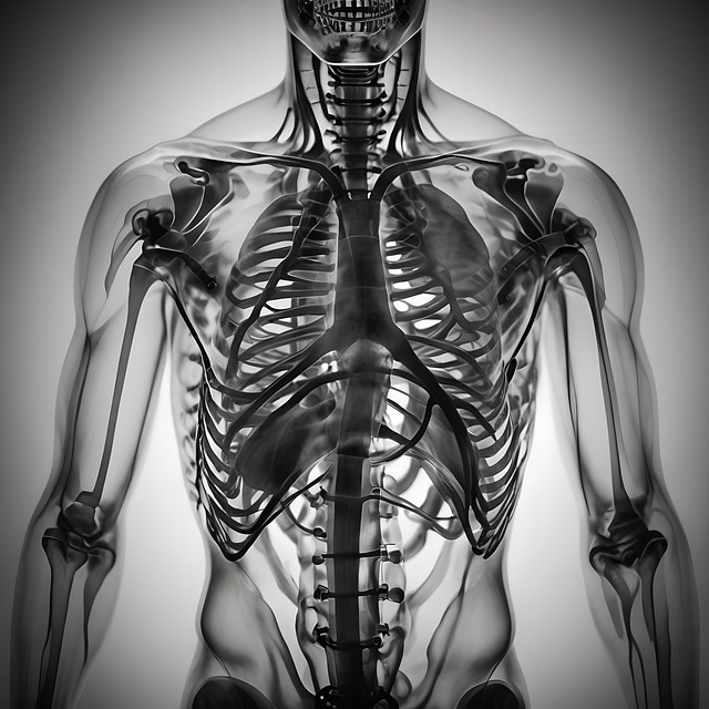
Fluoroscopy is a dynamic imaging technique providing real-time feedback on lung function, detecting abnormalities like collapsed lungs or air leaks. It complements thoracic MRI by visualizing lung movements continuously, enabling precise diagnosis and treatment planning. Future applications include advanced computing and hybrid imaging modalities combining fluoroscopy with thoracic MRI for enhanced lung condition evaluation and improved patient outcomes.
“Fluoroscopy emerges as a valuable tool in evaluating lung function, offering dynamic imaging capabilities that complement static methods like thoracic MRI. This article delves into the intricacies of fluoroscopy, exploring its role in assessing lung health and structure. We examine how this technology visualizes the lungs in real-time, highlighting its benefits and limitations compared to thoracic MRI. Furthermore, we discuss future advancements that may revolutionize lung imaging, shaping the next generation of respiratory care.”
Understanding Fluoroscopy: A Tool for Lung Assessment
Fluoroscopy is a dynamic imaging technique that plays a pivotal role in evaluating lung function, offering real-time visual feedback on respiratory dynamics. Unlike static imaging methods like thoracic MRI, fluoroscopy provides continuous insights into the movement of air within the lungs, making it invaluable for assessing breathing disorders and monitoring treatment responses.
By tracking the flow of contrast agents or breath-related changes, fluoroscopy can detect abnormalities in lung structure and function, such as collapsed lungs, air leaks, or narrowing of airways. This non-invasive approach allows healthcare professionals to make informed decisions, tailor treatments, and ultimately improve patient outcomes related to respiratory health.
How Fluoroscopy Works in Visualizing Lungs
Fluoroscopy is a specialized imaging technique that plays a pivotal role in evaluating lung function, often as an adjunct to other diagnostic tools like the more comprehensive thoracic MRI. Unlike static images, fluoroscopy provides dynamic, real-time visualization of internal structures, making it particularly useful for assessing the lungs’ intricate movements during respiration.
The process involves continuous X-ray imaging at a high frame rate, allowing doctors to observe the expansion and contraction of the lungs over time. This capability enables the detection of subtle abnormalities in lung structure or function that might be obscured by traditional imaging methods. By tracking the movement of air within the lung parenchyma, fluoroscopy helps identify issues such as air leakage, congestion, or restrictions, providing crucial insights for accurate diagnosis and treatment planning.
Benefits and Limitations Compared to Thoracic MRI
Fluoroscopy offers several advantages over Thoracic MRI when evaluating lung function. It provides real-time imaging, allowing for dynamic assessment of lung mechanics and detection of subtle abnormalities in ventilation or perfusion. Fluoroscopy is also non-invasive, making it a preferred choice for many patients, especially those with breathing difficulties who may find MRI scans uncomfortable or challenging due to claustrophobia. Additionally, fluoroscopy is more accessible and generally faster than Thoracic MRI, often requiring less preparation time.
However, Thoracic MRI has its own set of strengths. It offers high-resolution cross-sectional images of the lungs and adjacent structures without exposure to ionizing radiation, making it a safer alternative for repeated imaging over time. While fluoroscopy excels in real-time analysis, MRI provides more detailed anatomical information, aiding in the diagnosis of complex lung conditions. The choice between these two techniques ultimately depends on the clinical question, patient factors, and available resources.
Future Applications and Advancements in Lung Imaging
As technology advances, future applications of lung imaging are poised for significant growth. Fluoroscopy, with its real-time capabilities and versatility, will continue to play a pivotal role in this evolution. The integration of advanced computing power and artificial intelligence can enhance fluoroscopic techniques, allowing for more precise analysis of lung structure and function over time. This could lead to improved monitoring of respiratory diseases and the development of personalized treatment plans.
Additionally, there is potential for further exploration of hybrid imaging modalities, combining fluoroscopy with other advanced techniques like thoracic MRI. Such combinations may offer unprecedented depth and resolution in evaluating lung conditions, enabling healthcare professionals to make more informed decisions. These advancements promise to transform lung imaging into a dynamic field, improving patient outcomes and expanding our understanding of respiratory health.
Fluoroscopy emerges as a valuable tool for evaluating lung function, offering dynamic imaging capabilities that complement static methods like thoracic MRI. While each has its strengths and limitations, fluoroscopy excels in real-time visualization, making it crucial for assessing conditions requiring rapid intervention. As technology advances, future applications of fluoroscopy in lung imaging promise to enhance diagnostic precision and patient outcomes, potentially revolutionizing the way we understand and treat respiratory diseases.
