MRI with contrast revolutionizes medical imaging by enhancing soft tissue visibility through intravenously administered agents that interact with magnetic fields, improving diagnostic accuracy in detecting conditions like tumors, inflammation, and muscle degeneration by revealing subtle structural variations.
Discover how contrast media enhance MRI with contrast visualization of soft tissues, providing crucial insights for accurate diagnoses. This article explores the unique challenges posed by soft tissue structures and the opportunities presented by advanced MRI technology. We delve into the science behind contrast media, examining their role in improving image quality and enabling more precise interpretation of MRI scans. Learn how these agents enhance accuracy, aiding healthcare professionals in detecting abnormalities and guiding treatment decisions.
Understanding Soft Tissues: Challenges and Opportunities
Soft tissues, encompassing muscles, ligaments, and organs, are integral to our bodies’ structure and function. However, their intricate nature presents challenges in medical imaging. Traditional MRI techniques often struggle to differentiate soft tissues due to their similar signal properties, leading to blurred visuals and diagnostic difficulties. This is where the introduction of contrast media comes into play, offering a game-changer in enhancing soft tissue visualization.
Contrast agents, when administered intravenously during an MRI scan, interact with magnetic fields, resulting in distinct signal enhancements. This simple yet powerful technique allows radiologists to differentiate between various soft tissues, improving diagnostic accuracy. With MRI with contrast, subtle structural variations and abnormalities within these tissues become more apparent, opening up opportunities for early detection and precise diagnosis of conditions such as tumours, inflammation, or muscle degeneration.
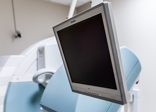
MRI Technology: A Non-Invasive Visualization Tool
Magnetic Resonance Imaging (MRI) has emerged as a powerful non-invasive visualization tool, offering unprecedented detail in imaging soft tissues. When combined with contrast media, MRI’s capabilities are further enhanced, enabling more precise and comprehensive analysis. Contrast agents, typically composed of small molecules or particles, are designed to interact with the body’s magnetic field differently than surrounding tissues. This interaction causes variations in signal intensity on the MRI scan, thereby highlighting specific structures and enhancing their visibility.
In an MRI with contrast, these agents can be injected into the bloodstream, allowing them to circulate throughout the body and accumulate in targeted areas. By strategically selecting contrast media with varying properties, radiologists can achieve better distinction between different types of soft tissues, such as muscles, organs, and tumors. This advanced visualization facilitates more accurate diagnosis, treatment planning, and monitoring of various medical conditions, making MRI with contrast an indispensable asset in modern healthcare.
The Role of Contrast Media in MRI Scans
Contrast media play a pivotal role in enhancing the visualization of soft tissues during Magnetic Resonance Imaging (MRI) scans. These substances, often administered intravenously, contain magnetic properties that differ from those of bodily tissues. When introduced into the body, they create distinct signals on the MRI scan, allowing for better distinction between soft structures like muscles, organs, and blood vessels. This improved contrast enables radiologists to interpret images more accurately, leading to precise diagnoses.
In MRI with contrast, these media not only highlight specific areas of interest but also help in detecting abnormalities or changes that might be subtle or difficult to discern using standard techniques. By providing a clearer picture of soft tissues, contrast media enhance the overall quality and reliability of MRI examinations, making them invaluable tools for clinical practice.
Enhancing Accuracy and Diagnosis with Contrast Agents
Contrast media play a pivotal role in enhancing the accuracy and diagnosis of soft tissue conditions through various imaging techniques, particularly in Magnetic Resonance Imaging (MRI). When administered to patients, these agents effectively differentiate between structures within the body, allowing radiologists to visualize soft tissues more clearly. By improving contrast, doctors can better identify abnormalities that might be difficult to discern without the use of contrast agents.
In the context of MRI with contrast, these agents are designed to interact with magnetic fields and radio waves, resulting in a marked increase in signal intensity on the final images. This enhanced contrast provides valuable insights into the microstructure and blood flow within soft tissues, aiding in the early detection and characterization of conditions such as tumors, inflammation, or injuries. The use of contrast media, therefore, significantly improves diagnostic accuracy, enabling healthcare professionals to make more informed decisions.
Contrast media play a pivotal role in enhancing the visualization of soft tissues through MRI scans, addressing many challenges encountered in conventional imaging. By improving signal contrast, these agents enable more accurate diagnosis and effective treatment planning. Incorporating MRI with contrast proves to be a powerful tool for healthcare professionals, offering unprecedented insights into soft tissue structures and dynamics.