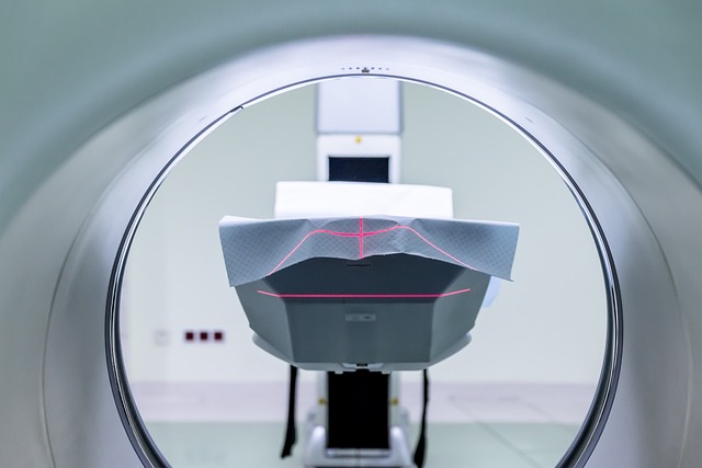
Traditional contrast agents like gadolinium chelates and iodine-based media enhance high-resolution MRI imaging by improving visualization of blood vessels, tissues with altered perfusion like tumors or inflamed areas, and soft tissues, enabling more accurate diagnoses in neurology and musculoskeletal radiology. Advanced MRI techniques, such as gradient echo sequences and 3D acquisitions, coupled with diverse contrast media tailored for specific applications, further elevate diagnostic accuracy while ensuring patient safety without ionizing radiation exposure.
“In the realm of medical imaging, Magnetic Resonance Imaging (MRI) stands as a cornerstone for detailed, non-invasive visualization. This article explores the diverse world of contrast media used in MRI, from traditional agents to cutting-edge innovations. We delve into the intricacies of high-resolution MRI imaging techniques, emphasizing their role in enhancing diagnostic accuracy. Additionally, safety concerns related to non-ionizing radiation are addressed, highlighting recent advancements in contrast media design for specific applications. By understanding these types and techniques, healthcare professionals can navigate the complex landscape of MRI.”
Traditional Contrast Agents in MRI
Traditional contrast agents play a vital role in enhancing specific structures or abnormalities within the body for high-resolution MRI imaging. These agents are designed to interact with magnetic fields, either by increasing signal intensity (positive contrast) or decreasing it (negative contrast), thus highlighting distinct anatomical features.
The most common types include gadolinium chelates, which are positively charged compounds that attach to water molecules, making them easier for the MRI machine to detect. This results in better visualization of blood vessels and tissues with altered perfusion, such as tumors or areas of inflammation. Other traditional agents, like contrast media containing iodine, are used for computed tomography (CT) scans but can also be utilized in specific MRI protocols to improve image quality and detection of abnormalities.
High-Resolution Imaging Techniques
High-resolution MRI imaging techniques play a pivotal role in enhancing diagnostic capabilities by providing detailed anatomical information. These advanced methods employ specialized contrast agents and sophisticated scanning strategies to produce crisp, clear images of internal structures. By focusing on subtle variations in tissue characteristics, high-resolution MRI enables radiologists to detect even the smallest abnormalities, leading to more accurate diagnoses and tailored treatment plans.
Among the various techniques, gradient echo sequences and three-dimensional (3D) acquisition methods stand out for their exceptional spatial resolution. Gradient echo sequences utilize magnetic field gradients to improve contrast, allowing for better differentiation between tissues based on their relaxation times. Meanwhile, 3D acquisitions capture detailed volumetric data, offering a comprehensive view of the body’s internal architecture. These high-resolution MRI imaging techniques have revolutionized medical diagnosis, especially in fields like neurology and musculoskeletal radiology, where minute changes can have significant clinical implications.
Non-Ionizing Radiation and Safety
One of the significant advantages of Magnetic Resonance Imaging (MRI) is its ability to provide detailed, high-resolution MRI imaging without exposing patients to ionizing radiation, a common concern with other diagnostic techniques like X-rays and CT scans. This feature makes MRI a safe choice for routine medical examinations and follow-up studies.
The absence of ionizing radiation ensures that patients are at minimal risk during the procedure. Contrast media, which enhances specific tissues or structures in an MRI scan, play a crucial role in achieving high-quality images without compromising safety. These media are carefully designed to interact with magnetic fields and provide contrast, allowing radiologists to differentiate between various types of soft tissues. By choosing appropriate contrast agents, healthcare professionals can optimize the benefits of MRI while maintaining patient safety, making it an indispensable tool for modern diagnostics.
Advanced Media for Specific Applications
With advancements in technology, various contrast media have been developed for specific applications in high-resolution MRI imaging. These advanced agents play a crucial role in enhancing the visibility of particular structures or abnormalities within the body, thereby improving diagnostic accuracy. For instance, gadolinium-based contrast agents are commonly used to highlight blood vessels and brain tissue, enabling radiologists to detect vascular malformations or tumors more effectively.
Additionally, specialized media designed for targeted imaging have emerged, allowing for more precise evaluations. These include agents that specifically bind to certain types of cells or tissues, such as those targeting cancerous cells or inflammatory sites. Such innovations in contrast media have not only elevated the quality of MRI examinations but also opened new possibilities for personalized and effective healthcare.
In conclusion, contrast media play a pivotal role in enhancing the diagnostic capabilities of MRI, particularly in achieving high-resolution MRI imaging. From traditional agents to advanced formulations tailored for specific applications, these substances continue to evolve, offering improved safety profiles and unparalleled visual detail. Understanding the diverse types of contrast media available is essential for healthcare professionals aiming to navigate the complex landscape of MRI technology and provide optimal patient care.
