Cancer diagnostic scans like CT, MRI, and PET scans use radiation to create detailed images of internal body structures, with modern technologies reducing dose levels. Healthcare professionals carefully weigh benefits against risks, selecting scan types based on patient needs and adhering to safety guidelines. Future advancements in tumor imaging include faster, lower-dose technologies like advanced MRI and CT scanners, along with AI algorithms for precise analysis and hybrid imaging techniques for comprehensive tumor understanding.
In the quest for early cancer detection, understanding radiation exposure risks associated with diagnostic scans is paramount. This article delves into the intricate details of how various cancer scans, including CT, MRI, and PET, impact patients’ health due to their radiation dose. We explore strategies to minimize these risks and highlight emerging trends in low-dose tumor imaging technologies, aiming to balance effective diagnosis with patient safety. By understanding these dynamics, healthcare professionals can make informed decisions, ensuring optimal tumor imaging while mitigating potential harm.
Understanding Radiation Exposure in Cancer Diagnostic Scans
Cancer diagnostic scans, including CT, MRI, and PET scans, utilize radiation to create detailed images of the body’s internal structures. Understanding the concept of radiation exposure is crucial when it comes to these scans as it helps in navigating the balance between the benefits of accurate tumor imaging and potential long-term health risks. The amount of radiation a patient receives during each scan is measured in millisieverts (mSv). While modern scanning technologies have significantly reduced radiation doses over the years, repeated or high-dose exposures may increase the risk of developing cancer later in life.
Healthcare professionals carefully consider the indications for each scan, ensuring that the potential benefits outweigh the risks. For example, PET scans, known for their ability to detect metabolic activity, often provide valuable information about tumor growth and treatment response. However, the higher radiation dose associated with PET scans requires careful planning and justification. By following guidelines and using advanced imaging techniques, medical experts can optimize tumor imaging while minimizing radiation exposure, balancing precision diagnostics with patient safety.
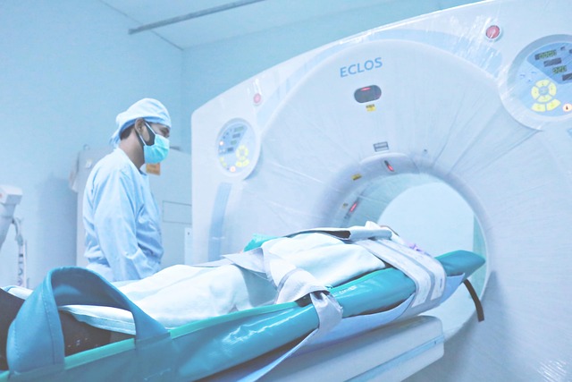
Types of Cancer Scans and Their Associated Risks
Cancer diagnostic scans, also known as tumor imaging, play a crucial role in early detection and treatment planning. However, it’s important to be aware of the associated risks, as these procedures involve exposure to radiation. Common types include X-rays, CT (computed tomography) scans, MRI (magnetic resonance imaging), and PET (positron emission tomography).
X-rays, often used for chest or dental imaging, deliver low doses of radiation but frequent exposure can be concerning. CT scans provide more detailed images but involve higher radiation levels. MRI uses strong magnetic fields and radio waves, making it a radiation-free option, yet lengthy procedures may increase anxiety. PET scans, while useful in certain cancers, also expose patients to radiation due to the radioactive tracers used for tumor detection. Understanding these risks is vital for informed decision-making regarding cancer diagnosis and treatment planning.
Minimizing Risk: Safety Measures and Best Practices
Minimizing risk is paramount when it comes to cancer diagnostic scans, as safety measures and best practices play a crucial role in protecting patients from unnecessary radiation exposure. Healthcare professionals are well-trained to utilize advanced technologies and techniques designed to optimize image quality while minimizing radiation doses. This includes selecting appropriate scan types tailored to specific symptoms or suspected conditions, as different imaging modalities emit varying levels of radiation. For instance, modern CT scanners employ faster acquisition times and dose-reducing protocols, significantly lowering the effective radiation dose compared to traditional machines.
Additionally, leveraging iterative image reconstruction algorithms enhances image quality without augmenting radiation exposure. These algorithms optimize scan parameters in real-time, allowing for adjustments to reduce dose while maintaining clear tumor imaging. Proper patient positioning and shielding are also essential practices. Patients should be educated on proper preparation, such as removing metal objects and ensuring they inform healthcare providers about any existing implants or devices. Healthcare staff must adhere to strict protocols, including regular calibration of equipment and adherence to recommended exposure limits for both patients and medical personnel.
Future Trends in Low-Dose Tumor Imaging Technologies
The future of cancer diagnosis looks promising with a growing focus on developing low-dose tumor imaging technologies. These innovations aim to revolutionize the way tumors are detected and monitored, minimizing radiation exposure while maintaining high diagnostic accuracy. Researchers and medical engineers are exploring advanced modalities such as magnetic resonance imaging (MRI) and computed tomography (CT) with optimized protocols to reduce scan times and dose levels without compromising image quality.
One of the key trends is the integration of artificial intelligence (AI) algorithms to analyze complex data sets from these scans, enabling more precise tumor characterization. AI-driven systems can detect subtle abnormalities and provide early indications of cancerous growths, potentially improving treatment outcomes. Additionally, hybrid imaging techniques that combine multiple modalities are being developed, offering a comprehensive view of tumors and their microenvironment, which could lead to personalized treatment strategies.
Cancer diagnostic scans, while crucial for early detection and treatment planning, carry a small but measurable risk due to radiation exposure. By understanding the types of scans, their associated risks, and implementing safety measures like those discussed in this article, patients and healthcare providers can make informed decisions. Future advancements in low-dose tumor imaging technologies hold promise for reducing these risks even further, paving the way for more frequent and effective cancer screenings. Ultimately, continued research and adoption of best practices in tumor imaging will contribute to better patient outcomes and enhanced quality of life during and after cancer treatment.