MRI techniques like Diffusion Tensor Imaging (DTI) offer powerful brain diagnosis tools, providing detailed insights into both brain structure (via DTI's mapping of neural tracts) and function (fMRI tracking brain activity). CT scans excel in rapid emergency assessments. PET scans monitor metabolic activities and neurotransmitters, aiding in conditions like Alzheimer's and neurological disorders. Together, these methods provide an unparalleled comprehensive view of the brain, assisting in accurate diagnosis and treatment planning for various conditions.
Medical imaging plays a pivotal role in brain diagnosis, offering a window into its intricate structure and function. This article explores four powerful techniques that revolutionize neurology. From Magnetic Resonance Imaging (MRI) revealing brain anatomy to Positron Emission Tomography (PET) mapping metabolic activity, each method provides unique insights. Computed Tomography (CT) scans offer swift assessments in emergencies, while Diffusion Tensor Imaging (DTI) specifically maps white matter tracks, enhancing our understanding of neural connectivity.
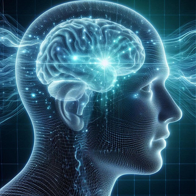
Magnetic Resonance Imaging (MRI): Unveiling Brain Structure and Function
Magnetic Resonance Imaging (MRI) is a powerful tool for brain diagnosis, offering detailed insights into both structure and function. This non-invasive technique employs strong magnetic fields and radio waves to generate images of the brain’s internal structures. One advanced form of MRI, known as diffusion tensor imaging (DTI), focuses on the movement of water molecules within the brain. By analyzing these movements, DTI can map out neural fiber tracts, providing crucial information about connectivity between different brain regions.
MRI’s versatility allows it to detect a range of conditions, from tumors and cysts that may alter brain anatomy, to neurological disorders affecting cognitive functions. For instance, in multiple sclerosis, MRI scans can reveal changes in the white matter, indicative of damaged nerve fibers. Additionally, functional MRI (fMRI) measures brain activity by detecting blood flow changes, enabling researchers to identify areas activated during specific tasks or states, such as language processing or emotional responses.
Computed Tomography (CT) Scans: Rapid Assessment for Emergency Situations
In emergency situations where rapid assessment is crucial, Computed Tomography (CT) scans play a pivotal role in brain diagnosis. CT scanners use X-rays to create detailed cross-sectional images of the brain, allowing healthcare professionals to quickly identify bleeding, tumors, or structural abnormalities. This non-invasive technique provides valuable insights into the brain’s anatomy, making it an indispensable tool for initial evaluations, especially in critical care settings.
While CT scans offer a broad overview and are highly effective for acute conditions, they don’t capture the complex connectivity of neural pathways revealed by diffusion tensor imaging (DTI). DTI is a specialized form of MRI that tracks the movement of water molecules within brain tissue, providing detailed information about white matter tracts and neural communication. This advanced imaging technique complements CT scans, offering a more comprehensive understanding of brain structure and function, particularly in cases where subtle changes or neurological disorders are suspected.
Positron Emission Tomography (PET): Tracking Metabolism and Neurotransmitter Activity
Positron Emission Tomography (PET) is a powerful tool in brain diagnosis, offering unique insights into metabolic and neurotransmitter activities. This imaging technique tracks the movement of substances like glucose, which is vital for understanding how different areas of the brain function. By measuring changes in metabolism, PET scans can reveal active regions involved in cognitive processes, memory formation, or even conditions like Alzheimer’s disease.
Furthermore, PET scans can detect the presence and distribution of specific neurotransmitters, such as dopamine and serotonin. This capability is particularly beneficial for evaluating neurological disorders and psychiatric conditions, where disruptions in neurotransmitter systems play a significant role. When combined with other imaging methods like diffusion tensor imaging (DTI), PET provides a comprehensive view, linking brain structure to its chemical activity.
Diffusion Tensor Imaging (DTI): Mapping White Matter Tracks in the Brain
Diffusion Tensor Imaging (DTI) is a powerful tool in medical imaging that specifically focuses on mapping white matter tracks in the brain. By measuring the movement and direction of water molecules, DTI creates detailed 3D maps of neural pathways, providing crucial insights into the structure and integrity of the brain. This non-invasive technique allows healthcare professionals to visualize and quantify the connectivity between different brain regions, which is essential for accurate diagnosis and understanding neurological conditions.
DTI has become an invaluable resource in neurology and neurosurgery, enabling doctors to assess a range of disorders, including stroke, traumatic brain injuries, multiple sclerosis, and even normal aging. The detailed maps generated by DTI help identify lesions, demyelination, or changes in fiber tracts, aiding in the early detection and differentiation of various brain conditions. This advanced imaging method offers a unique perspective on brain function and structure, enhancing diagnostic accuracy and guiding treatment decisions.
In conclusion, medical imaging plays an indispensable role in brain diagnosis, offering diverse techniques such as MRI, CT scans, PET, and DTI. Each method provides unique insights into brain structure, function, metabolism, and connectivity, enabling healthcare professionals to make accurate diagnoses and develop personalized treatment plans. Diffusion tensor imaging (DTI) stands out for its ability to map white matter tracks, enhancing our understanding of neural connections and contributing significantly to neuroscientific research and clinical practice.